

What a Dental CBCT Scan Can Reveal: Carotid Artery Findings, Hidden Infections, and Radiation Facts
We take a CBCT — a 3D dental scan — to plan an implant, evaluate a tricky root, or map out someone's sinus and jawbone anatomy. Most of the time, we find exactly what we went looking for.
But often, the scan shows something we weren't specifically looking for at all: a calcified spot near a carotid artery, or an area of bone change at a tooth root that never caused a moment of pain. Those can be serious findings. Here's more on why, and what it means if one shows up in your own CBCT scan.

Key takeaways
- A dental CBCT can show a possible carotid artery calcification — found in roughly 6 to 12% of scans, depending on the population studied.
- What it can do is show a calcification in the expected carotid region that may be consistent with calcified atherosclerotic plaque. It cannot confirm the diagnosis, measure blood flow, or tell us how narrowed the artery may be — which is why medical follow-up matters.
- Effective radiation dose from a CBCT varies substantially with field of view, patient size, and protocol. We choose the smallest field and lowest exposure that will reliably answer the clinical question.
- We review the entire volume of every scan, not just the region of interest we were asked to look at, because we often find other medically relevant things worth knowing about.
- We look actively for connections between your mouth and the rest of your health, and we bring your physician into the conversation when something matters.
In this article
What is a dental CBCT scan?
CBCT stands for cone beam computed tomography. The machine rotates around you, capturing many individual images with a cone-shaped X-ray beam, and software stitches them into a three-dimensional model of the area.
A standard dental X-ray is still a fine tool, but it flattens three-dimensional anatomy onto a flat image, so roots, bone, and restorations overlap and hide one another. CBCT lets us move through the anatomy one thin slice at a time, from any angle. Sometimes that changes what we can see entirely.
We don't order a CBCT for every patient or every problem. It carries more radiation than a standard X-ray, so we use it selectively — when the extra three-dimensional detail is likely to change a diagnosis or a treatment plan. That's the same principle the American Association of Endodontists and American Academy of Oral and Maxillofacial Radiology reaffirmed in their 2025 joint update, released January 2026: use CBCT deliberately, not routinely.1
Carotid artery calcification on a dental scan
Depending on the field of view, part of your neck and the expected path of your carotid arteries falls inside the frame. In some patients, we see areas of calcification in that region that may be consistent with calcified atherosclerotic plaque — deposits associated with atherosclerosis, the slow buildup of plaque inside artery walls.
This isn't rare, and it isn't trivial.
Those middle and right figures come from a prospective study out of Umeå University in Sweden, which followed patients who had this finding on routine dental imaging for a mean of 9.5 years and tracked myocardial infarction, stroke, and vascular death.3 Those are the kinds of numbers that make us look carefully, every single time.
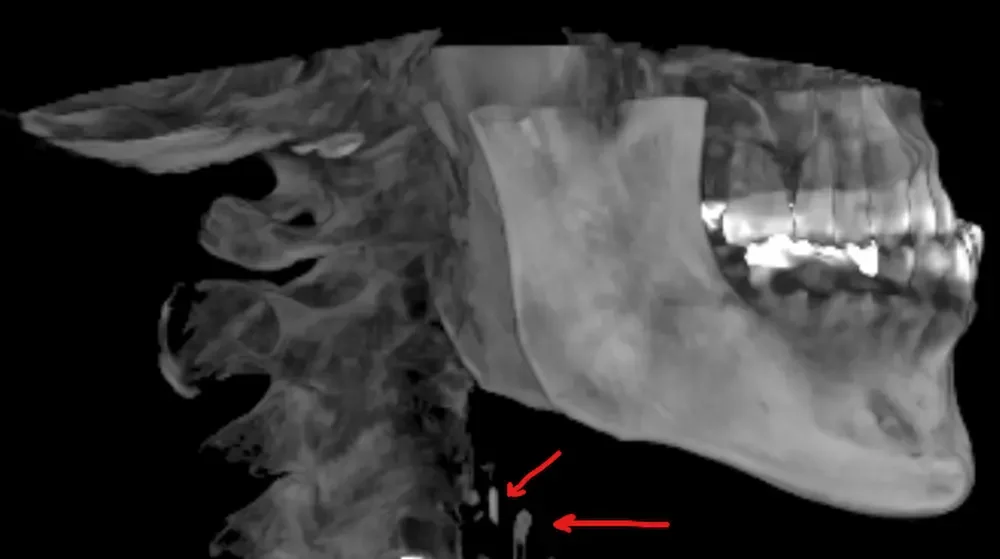

Does that mean an artery is blocked?
Not necessarily — and that distinction is worth understanding, because it's exactly what makes this finding useful rather than alarming. Calcification and narrowing aren't the same thing. Arteries remodel outward as plaque accumulates, so a calcified vessel can still carry normal blood flow.
What a CBCT can do is show a calcification in the expected carotid region that may be consistent with calcified atherosclerotic plaque. It cannot confirm the diagnosis, measure blood flow, or tell us how narrowed the artery may be — which is why medical follow-up matters. The logic is similar to coronary artery calcium scoring in cardiology: that test doesn't measure blockage either. It reflects plaque burden, and it remains one of the better-validated risk tools cardiologists use.
Most routine wellness visits don't include imaging of the neck. Ours sometimes does.
So the answer is layered: it doesn't mean your artery is blocked, and we can't diagnose blockage from a dental scan. But it does mean something worth following up on — a signal your physician can evaluate that they would otherwise have no particular reason to look for. If this were our own family member, we'd want someone to point it out plainly, and we'd want them to take the next step.
Why we read every scan whole
A CBCT captures far more than the tooth or implant site that sent us looking. When we scan to plan a dental implant, the volume also contains jawbone, sinuses, airway, joints, and — depending on the field of view — the vessels of the neck.
The American Academy of Oral and Maxillofacial Radiology addressed this directly in its executive opinion statement on CBCT: practitioners are expected to review the whole exposed volume, not just the region they ordered the scan for. The statement notes that oral and maxillofacial radiologists can assist practitioners who are unwilling to accept that responsibility.4 We accept it. Here's what that looks like in practice:
- We review the full volume. Every scan, every time — the region of interest and everything else the scan captured.
- We document what we find. Findings go in your chart in plain language, whether they're dental, incidental, or simply worth watching.
- We show you the images. On the screen, walking through what we're seeing, so you understand your own anatomy rather than taking our word for it.
- We loop in your physician when it matters. If something belongs in a medical conversation, we make sure it gets there instead of sitting in a dental file.
There are practical reasons this matters beyond curiosity. Patients preparing for joint replacement are often asked to have a dental evaluation beforehand — a check for active infection a surgeon would rather address before, not after, a new hip or knee goes in. The AAOS's 2024 clinical practice guideline reviews the evidence on dental screening and antibiotic prophylaxis around arthroplasty and is worth reading with your surgeon, since practice varies.5 When we already have a CBCT in hand for another reason, checking it for hidden sources of infection costs nothing extra and may answer your surgeon's question before they ask it.
A dental infection may remain localized and painless for a long time, but it still deserves evaluation. Full-volume review means carefully evaluating every structure captured by the scan — a habit we build into every case because we're looking for connections to the rest of your health, not just the reason you're in the chair.
How much radiation is involved?
Less than most people expect when they hear "3D CT scan" — but the honest answer is that it depends, and we'd rather give you the real range than a comforting single number.
Effective dose varies substantially based on the field of view, patient size, and imaging protocol. Published values for the Planmeca ProMax 3D Mid range from very low-dose scans of roughly 4 µSv up to approximately 190 µSv for certain larger standard protocols.6 Some low-dose scans are comparable to a long flight; larger scans involve more radiation. We choose the smallest field of view and lowest exposure that will reliably answer the clinical question — the ALARA principle (As Low As Reasonably Achievable) that governs how every dentist and physician should think about imaging.
For context, in microsieverts (µSv), the standard unit of effective dose:
| Source | Typical effective dose |
|---|---|
| Single digital bitewing or periapical X-ray | 1–5 µSv |
| Lateral cephalometric X-ray | 1–6 µSv |
| Digital panoramic X-ray | 15–20 µSv |
| CBCT, Planmeca ProMax 3D Mid (varies by protocol) | ~4–190 µSv |
| Round-trip transatlantic flight | ~40 µSv |
| Medical CT scan (head, chest, or abdomen) | 500–10,000+ µSv |
| Natural background radiation, per year (US average) | ~3,000 µSv |
Planmeca's published dose table for this unit lists a low-dose protocol as low as 4 µSv for a small sinus view, and 190.7 µSv for a 16 × 16 cm wide-view, high detail scan.6 Even at the upper end, that remains a fraction of a single medical CT scan and well within the range of ordinary annual background exposure. The FDA's guidance on dental cone-beam CT is a good plain-language resource if you'd like to read more.7
The oral-systemic connection
Here's the real reason we care when your gums bleed a little at your cleaning, or an X-ray shows a few millimeters of bone loss: it was never really about the gums. Chronic periodontal inflammation doesn't stay put. The same bacteria and inflammatory signaling living under an infected gumline show up in the bloodstream, in blood vessels, in the same inflammatory pathways implicated in heart disease and poorly controlled diabetes.
This isn't a fringe idea. In 2020, the European Federation of Periodontology and the World Heart Federation convened a joint consensus workshop specifically on this relationship, and concluded there is strong epidemiological evidence that periodontitis is associated with increased risk of future cardiovascular disease — recommending that patients with periodontitis actually be told so.8 Cardiologists and periodontists sat in the same room to write that. A bleeding gum, by itself, tells us almost nothing interesting. What it's usually a sign of is what makes us pay attention.
If it were only about a bleeding gum, we honestly wouldn't be talking about it.
We want to be precise about what the evidence supports, because precision is what makes this credible instead of a talking point. The research shows a real, consistent association between periodontal disease and cardiovascular disease — not proof that one causes the other, and not proof that treating your gums will prevent a heart attack. Nobody can honestly promise that, and we won't. But "just an association" undersells it badly. Associations this consistent, across this much research, are exactly the kind of signal that changes how a careful clinician practices.
So here's what we'll actually tell you: if it were our own mother with bleeding gums, we wouldn't stop at "floss more." We'd want her to know that inflammation sitting under her gumline doesn't stay under her gumline, and that treating it is one of the few genuinely low-risk, high-value things she could do — not just for her smile, but for her whole-body health.
- Periodontal disease is a chronic inflammatory condition, and inflammation doesn't respect the border between your mouth and the rest of you.
- Dental findings deserve evaluation even when they don't hurt, because "no pain" doesn't mean "no systemic signal."
- Diabetes, smoking, nutrition, dry mouth, airway and breathing patterns, and overall health all shape your dental risk — and your dental health shapes them right back.
- Unexpected imaging findings belong in a conversation with you and your physician, not filed away.
- We coordinate with physicians whenever a finding crosses the line between oral and medical health, because your care shouldn't live in silos.
To us, whole-body dentistry means treating a bleeding gum like the systemic signal it actually is — without overselling what we can promise, and without pretending it's "just" a dental problem either. If that's how you think about your own health, you'll probably feel at home here.
When we recommend a CBCT
At Teuscher Legacy Dental, a CBCT might come up for:
- Dental implant planning
- Complex extractions and impacted wisdom teeth
- Bone graft and socket preservation planning
- Unclear endodontic symptoms, or evaluating a previously treated root canal
- Suspected root fracture or resorption
- Complex anatomy near a sinus or nerve
- Selected airway and orthodontic or TMJ evaluations
- Any other situation where 3D information is likely to change your care
We don't recommend advanced imaging simply because we own the technology. The scan has to answer a real question. When it's appropriate, we'll show you the images, walk you through what we see in plain language, and tell you if something belongs in a conversation with your physician. You can see more of the services we offer or read answers to common questions.
Periapical findings that don't hurt
A CBCT is also useful for a tooth with unclear symptoms, or a previously completed root canal. A tooth can carry inflammation around the root tip without causing pain, swelling, or any obvious sign. On imaging, this shows up as a periapical lesion — an area of bone change at the tip of the root that may reflect apical inflammation or infection. A standard X-ray can miss these entirely when overlapping roots or dense bone get in the way.
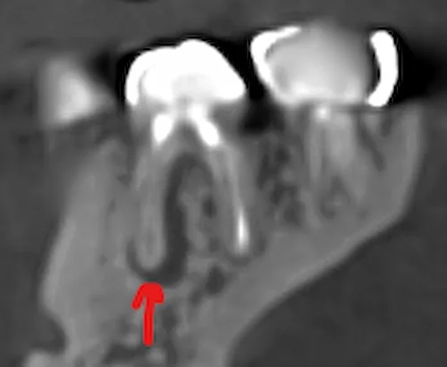
A dark area on a CBCT doesn't automatically mean the tooth has to come out. We weigh it against your history, symptoms, clinical testing, conventional X-rays, restorability, and long-term prognosis. Depending on the situation, the right next step might be monitoring, a root canal, retreatment, a referral to an endodontist, apical surgery, or — only when the tooth truly can't be saved — extraction with bone grafting.
Better imaging doesn't replace judgment. It gives us better information to use it with.
Want a dental team that looks at the whole picture?
We serve St. Charles, Campton Hills, Geneva, Batavia, Elburn, South Elgin, Wasco, Wayne, and the surrounding Fox Valley. New patients welcome.
Schedule a visit Ask us a questionFrequently asked questions
Can a dental scan detect blocked arteries?
No. A dental CBCT can show a calcification in the expected carotid region that may be consistent with calcified atherosclerotic plaque. It cannot confirm the diagnosis, measure blood flow, or tell us how narrowed the artery may be — which is why medical follow-up matters.
Is carotid artery calcification the same as a coronary calcium score?
No, though the logic is similar. Both reflect plaque burden rather than blockage. A coronary calcium score comes from a dedicated medical CT scan and produces a validated numerical risk score; a dental CBCT finding is an observation that warrants medical follow-up.
How much radiation does a dental CBCT involve?
Effective dose varies substantially with field of view, patient size, and protocol. Published values for the Planmeca ProMax 3D Mid range from roughly 4 µSv for a small low-dose scan up to about 190 µSv for certain larger standard protocols — a fraction of a medical CT scan (500 to several thousand µSv).
Do dentists review the whole CBCT scan or just the tooth?
The American Academy of Oral and Maxillofacial Radiology expects practitioners to review the entire exposed volume, not only the region of interest. That's our standard at Teuscher Legacy Dental — every scan, every time.
Should I see a dentist before joint replacement surgery?
Many orthopedic surgeons ask for a dental evaluation beforehand. The AAOS published an updated clinical practice guideline in 2024 addressing dental screening and antibiotic prophylaxis around joint replacement; it's worth reviewing with your surgeon. If yours has asked, we're glad to help.
Can a tooth have a problem without hurting?
Yes. A tooth with a dead nerve, a previous root canal, or chronic low-grade inflammation around the root can cause little or no pain. No pain doesn't always mean a healthy tooth.
Does everyone need a dental CBCT scan?
No. We use CBCT selectively, when the added 3D information is likely to improve diagnosis or treatment planning. Conventional dental X-rays remain appropriate for most routine care.
Clinical sources
- Special Committee to Revise the Joint AAE/AAOMR Position Statement. AAE and AAOMR Joint Position Statement: Use of Cone-Beam Computed Tomography in Endodontics — 2025 Update. Oral Surgery, Oral Medicine, Oral Pathology and Oral Radiology. 2025.
- Dief S, Veitz-Keenan A, Amintavakoli N, McGowan R. A systematic review on incidental findings in cone beam computed tomography (CBCT) scans. Dentomaxillofacial Radiology. 2019;48(7):20180396. doi:10.1259/dmfr.20180396
- Garoff M, Ahlqvist J, Edin LT, Jensen S, Levring Jäghagen E, Petäjäniemi F, Wester P, Johansson E. Bilateral vessel-outlining carotid artery calcifications in panoramic radiographs: an independent risk marker for vascular events. BMC Cardiovascular Disorders. 2019;19(1):225. doi:10.1186/s12872-019-1211-3
- Carter L, Farman AG, Geist J, Scarfe WC, Angelopoulos C, Nair MK, Hildebolt CF, Tyndall D, Shrout M. American Academy of Oral and Maxillofacial Radiology executive opinion statement on performing and interpreting diagnostic cone beam computed tomography. Oral Surgery, Oral Medicine, Oral Pathology, Oral Radiology and Endodontology. 2008;106(4):561-562.
- American Academy of Orthopaedic Surgeons. Prevention of Total Hip and Knee Arthroplasty Periprosthetic Joint Infection in Patients Undergoing Dental Procedures: Evidence-Based Clinical Practice Guideline. Published November 18, 2024.
- Planmeca. Planmeca ProMax 3D Mid — Effective Doses (ICRP 1990 and ICRP 2007). Manufacturer dosimetry table.
- U.S. Food and Drug Administration. Dental Cone-Beam Computed Tomography.
- Sanz M, Marco del Castillo A, Jepsen S, et al. Periodontitis and cardiovascular diseases: Consensus report of the joint workshop of the European Federation of Periodontology and the World Heart Federation. Journal of Clinical Periodontology. 2020;47(3):268-288. doi:10.1111/jcpe.13189
This article is intended for patient education and does not replace an individualized dental or medical evaluation. Dental CBCT imaging may reveal findings that warrant medical follow-up, but it does not diagnose cardiovascular disease or any other medical condition.