

What Should a Bone Graft Look Like 1 Month After Surgery?
Real photos & honest healing expectations — from a surgical case at Teuscher Legacy Dental in St. Charles, IL
Patients Ask Us:
“Is what I’m seeing one month after my extraction and bone graft normal? Is it supposed to look like that?”
About the author
This article was written by Dr. Brayden Teuscher, a general dentist with a surgical focus on tooth extractions, bone grafting, and dental implants in St. Charles, IL, serving Kane County, Campton Hills, Geneva, Elburn, and surrounding communities.
At Teuscher Legacy Dental, we place extraction socket preservation bone grafts every week. The 1-month mark can be a major milestone — usually, soft tissues have mostly healed, bone formation is continuing under the surface, and discomfort should be minimal. But patients often aren’t sure what “normal” healing looks like at this stage.
In this post, I’ll walk you through:
What normal 1-month bone graft healing should look like
Real photos from a current patient case
What we evaluate clinically at this stage
When you might need to be seen sooner
Case Overview: Why This Bone Graft Was Done
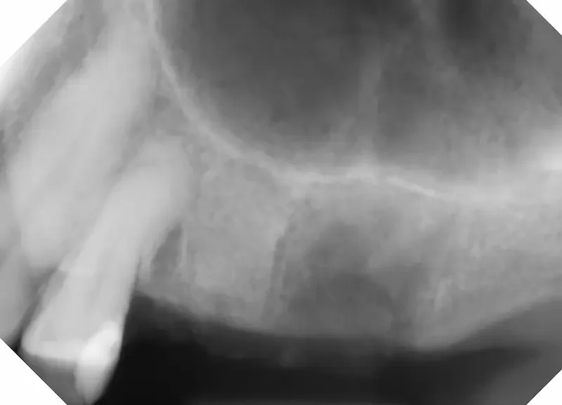
Tooth #13 (center of X Ray) had an old root canal that was infected. In the 2D X Ray, the infection is difficult to see.

The CBCT 3D slice clearly shows the dark infection around the edge of the root. To the right side, a previous extraction site not done in our office.
This patient is a female in her 60s. In the 2D X Ray above, the tooth #13 doesn’t seem so bad. But in the 3D CBCT scan to the right, we can clearly see an infection around the root from the old root canal. This patient did not want to re-do the root canal, and preferred to have the tooth extracted and socket preservation bone graft placed to prepare for future dental implant.
*Notes for this case: The CBCT imaging on the right allows us to see things we never would have known about from a 2D X Ray. Notice how clearly you can see the abscess in the CBCT slice. CBCT imaging in dentistry is the standard of care for implant planning, and an incredibly powerful diagnostic tool when evaluating for infection and other conditions.
Also, This patient had an infected tooth #14 extracted and a collagen base bone graft placed at another office about 2 months before seeing us. That is the darkness on the right side of the X Ray and CBCT screenshot above. This type of bone graft heals more slowly and often less predictably than the demineralized bone particles we most often use. I did not take a picture of this particular site that day since we were evaluating the tooth #13 in front of it. At our office, we use that same collagen base graft in certain select situations. That isn’t the point of this particular post, but worth noting.
The Extraction and Socket Preservation Procedure Day
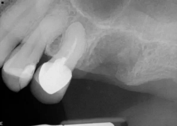
After the tooth was extracted and bone graft placed. Notice how bone particles fill the previous root and infection space.

Photo of the collagen plug and white sutures over the bone particle graft. You can see some of the bone particles over the top of the collagen. It is normal for some of the particles to spill out at first.
In the X Ray above you can see the tooth socket filled with bone graft material. (Our graft is center of X Ray, right next to the tooth that is still there. To the right of center is a larger infection that was previously grafted at a different office using different materials.) In the photo, You can see the pink collagen plug, white sutures that criss-cross over the top, and even some gray bone particles that spilled out onto the collagen. This is normal and the grafted site looks great!
Our method of using demineralized particulate bone with a collagen plug and sutures over the top helps by:
Preserving alveolar ridge (jaw bone) width and height
Supporting new bone formation
Protecting the graft site while soft tissue heals
Extraction and bone graft procedures are quite routine at our office, and this case is a good example of what happens.
What Real Healing Looks Like at 1 Month

One month after procedure, the pink gum tissue is almost fully closed. The darker red area is immature gum tissue. The light pink on the outside is healthy. If it was infected it would be much redder and more swollen.
One month later, our patient was doing great. In general, the way a bone graft site looks after one month can vary dramatically based on patient factors like age, medical history, current medications, smoking status, and the extent of tooth infection prior to extraction. But this picture should give you a good idea of what to expect.
At 1 Month, we see Soft Tissue Progress
By 4 weeks after surgery you should see:
Gum tissue mostly closed over the grafted site
A smooth contour of healed soft tissue
Little to no visible crater
Minimal redness or swelling
This means the gum has largely healed on the surface and your body is moving into deeper phases of bone rebuilding.
Clinically, this is great healing. If the soft tissue border looks pink and stable without irritation, that’s exactly what we want to see.
Bonus: What An Extraction and Bone Graft Site Should Look Like:
What’s Happening Under the Surface at 1 Month
Even if it “looks all the way healed,” real bone growth is still happening deep inside:
The graft material is acting as a scaffold for new bone cells
True bone mineralization continues for months, not days
At 1 month, early bone formation is already underway but will continue strengthening
This matches typical bone graft healing timelines described by peer reviewed surgical literature, where bone integration usually spans weeks 2–6 and beyond.
So if the site seems flatter, closed, or even slightly irregular — that’s normal. The surface has healed; the bone is still maturing.
How It ShouldFeel at 1 Month
Most patients report:
Little to no discomfort
Occasional slight pressure or tightness
More confidence chewing elsewhere — and not thinking about the graft site
By this stage, most pain has resolved and you should feel pretty normal. Some sensitivity is okay, but sharp or worsening pain isn’t. Increasing pain or swelling in that area can indicate infection.
When Healing Isn’t Typical
Contact your dentist if you notice:
Increasing pain or throbbing
Swelling that worsens after initial improvement
Foul taste, active drainage, or pus
Fever or feeling unwell
These are not typical at one month and may need evaluation.
Final Takeaway
At 1 month after a bone graft, your site should look healed on the surface, feel comfortable, and show at least some pink, stable gum tissue.
Deeper bone healing is still in progress — and that’s exactly how it should be.
Healing isn’t “instant perfection” — it’s progress. If you’re not sure whether your graft is healing normally, we can help evaluate and reassure you.
Have Questions or Want Your Site Evaluated?
Call us at (630) 762-0000 or request a surgical evaluation at Teuscher Legacy Dental in St. Charles, IL.
Healing isn’t always pretty — but knowing what’s normal makes it a lot less stressful.
— Dr. Brayden Teuscher