

What Does an Extraction & Bone Graft Look Like 2 Weeks After Surgery?
Real Photos & X-Rays From a Surgical Case in St. Charles, IL
One of the most common questions we hear from patients after a tooth extraction and bone graft is:
“Is this what a bone graft is supposed to look like at 2 weeks?”
At Teuscher Legacy Dental in St. Charles, IL, we perform extractions and bone grafts all the time, and the 2-week mark can be a critical checkpoint. It’s often when the slowest healing patients feel better — but still worry that something doesn’t look “right”.
In this post, I’ll walk you through:
What normal healing looks like 2 weeks after extraction and bone graft
Real before-and-after photos and X-rays
What we evaluate clinically at this stage
When we’re reassured — and when we’re concerned
This is a real case from our surgical practice, not a stock example.
About the author
This article was written by Dr. Brayden Teuscher, a general dentist with a surgical focus on tooth extractions, bone grafting, and dental implants in St. Charles, IL, serving Kane County, Campton Hills, Geneva, Elburn, and surrounding communities.
Case Overview: Why This Tooth Needed Extraction & Bone Grafting
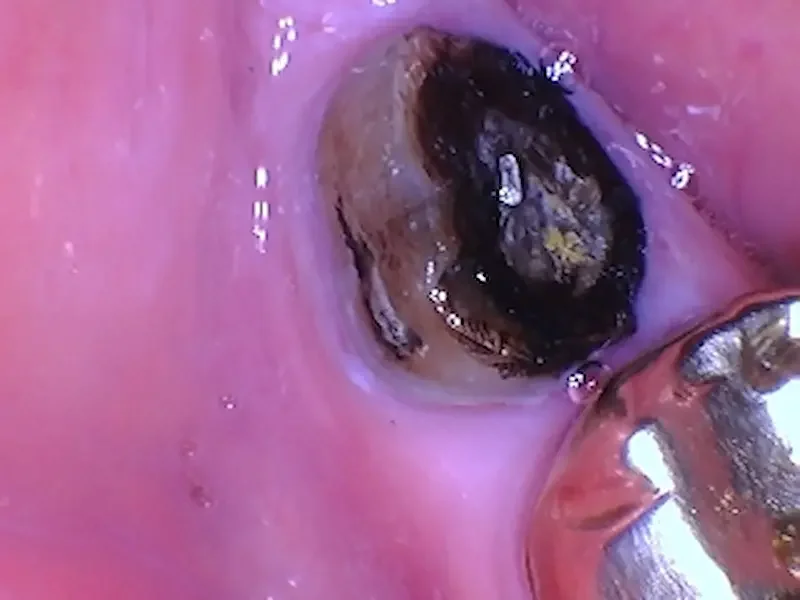
A lower right molar crown fell out. The darkness is mostly stain, but there are small areas of decay that likely contributed to the crown loosening. The tongue side of the remaining tooth is flush with the gums.
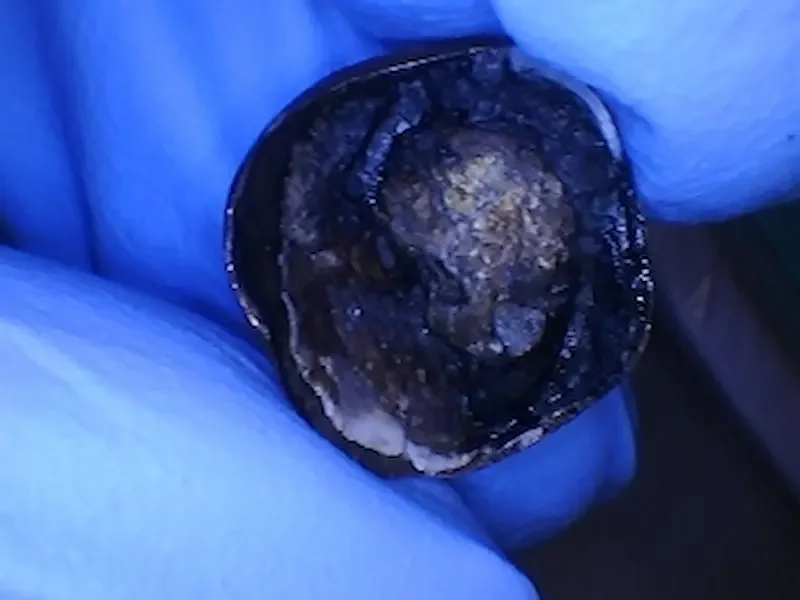
The inside of the crown that fell out. Some old cement came out with the crown.

An X-Ray of tooth #31 with crown missing. You can see this tooth had a previous root canal and large post in the back root.
This patient is a female in her 60s. She patient decided to have this tooth removed due to structural compromise that made long-term restoration unpredictable. Not everyone decides to get their tooth out, but this patient did. Because the patient is planning for a future dental implant, a socket preservation procedure with a bone graft was performed at the time of extraction.
Surgical goals of the bone graft:
Prevent collapse of the socket during early healing
Create a stable foundation for future implant placement
This was a routine procedure for us at our surgical practice. We extracted the tooth, then filled the root spaces with a 70/30 ratio cancellous to cortical cadaver bone particulate graft. Next we stabilized the graft with a collagen plug and suture to support predictable bone regeneration. Photos below:
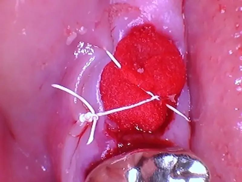
A photo of the grafted site. The red part is the collagen plug over the top of the particle graft. In this case one figure 8 suture was enough to keep everything stable for healing.
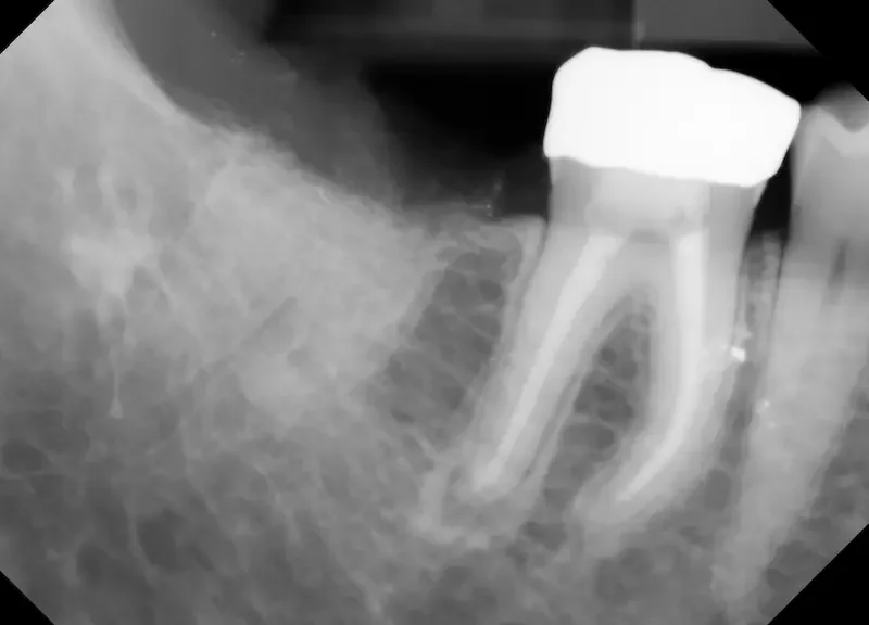
An X-Ray of the extraction site after bone graft placement. You can see how the lighter shade of the grafted particles fills in the space where the tooth roots were before the extraction.
What We Look for Clinically at the 2-Week Healing Mark
At two weeks, bone has not yet fully regenerated — and that’s important to understand.
This phase is primarily about soft tissue stability and graft protection.
At 2 weeks, we evaluate:
Whether the graft material has remained stable (versus completely fallen out)
How the gum tissue is closing over the site
Signs of inflammation or infection versus healthy healing
Whether the site is protected enough to continue healing undisturbed
This is often the visit where patients expect the site to “look filled in.” That usually does not happen yet — and that’s normal.
Real Photo: What You’re Seeing at 2 Weeks
2 Weeks After Surgery

The grafted site is healing well. The white and yellow granules toward the top of the photo are residual bone graft particles, not an infection. and toward the bottom near the gold crown you can see the gums are already almost fully closed over.
Our patient was concerned about the “yellow spots”. We explained these are particles from the bone graft, NOT an infection. They will likely exfoliate (fall out) on their own, and there is plenty of bone graft underneath them. It is very normal to lose a few of the graft granules, especially the cortical granules which are the bigger and denser particles in the particular mix we used in this case.
A big key that you can see in the photo is that the gum tissue around the site and in the cheek area was pink, and minimally swollen. If an infection were present, we’d see deep red, irritated or visibly swollen tissue. Usually it would be painful. Our patient had no pain.
Some of the gums closer to the gold crown are completely covering the extraction site already, which is great healing for someone in their 60s! Remember that medical conditions can affect healing timelines, so sometimes even for a younger person, healing this good doesnt happen for another week or two.
To sumarize:
The gum tissue has migrated inward and downward, almost fully closing the extraction socket
The socket appears shallower, but not closed
Mild surface irregularity is expected
This is normal healing.
True and final bone maturation occurs months later, not weeks.
Is It Normal for a Bone Graft to Look Hollow at 2 Weeks?
Yes — and this is one of the biggest misconceptions we get asked about.
At two weeks:
The body is still forming early connective tissue
New bone has not fully mineralized yet
The graft is acting as a scaffold, not final bone
If the site looked “fully filled in” at two weeks, that would actually be unusual.
Bonus content: What a bone graft should look like after 1 week healing.
Should There Still Be Discomfort at 2 Weeks?
Most patients report:
Minimal soreness or pressure
No sharp or worsening pain
Improved chewing comfort, especially on the opposite side
Pain that increases after week one is not typical and should be evaluated.
How We Know This Graft Is Healing Normally
From a surgical standpoint, this site shows:
Stable graft material
Healthy soft tissue color and contour
No signs of infection or graft loss
At this point, we’re looking for progression, not perfection.
Warning Signs That Are Not Normal at 2 Weeks
While most bone grafts heal uneventfully, contact your dentist if you notice:
Increasing pain after initial improvement
Persistent swelling or pressure
Foul taste or drainage
Fever or systemic symptoms
Early evaluation can prevent larger complications. When these symptoms are present, an infection is possible.
What Happens Next in the Healing Timeline?
Typical progression:
Weeks 0–2: Soft tissue stabilization
Weeks 3–6: Early bone formation begins
3–6 months: Bone matures and strengthens
Implant planning: Based on graft size and location
Every case is different — this is why follow-up matters. At our practice, each surgical case is unique and patients get custom treatment timelines depending on their unique needs.
Unsure If Your Bone Graft Is Healing Normally?
Many patients come to us worried — and leave relieved after we review healing clinically and on X-ray.
If you’ve had an extraction or bone graft and:
Something doesn’t look right
Healing feels slower than expected
You want a second opinion before implant placement
We’re happy to evaluate your site and give you a clear, honest assessment.